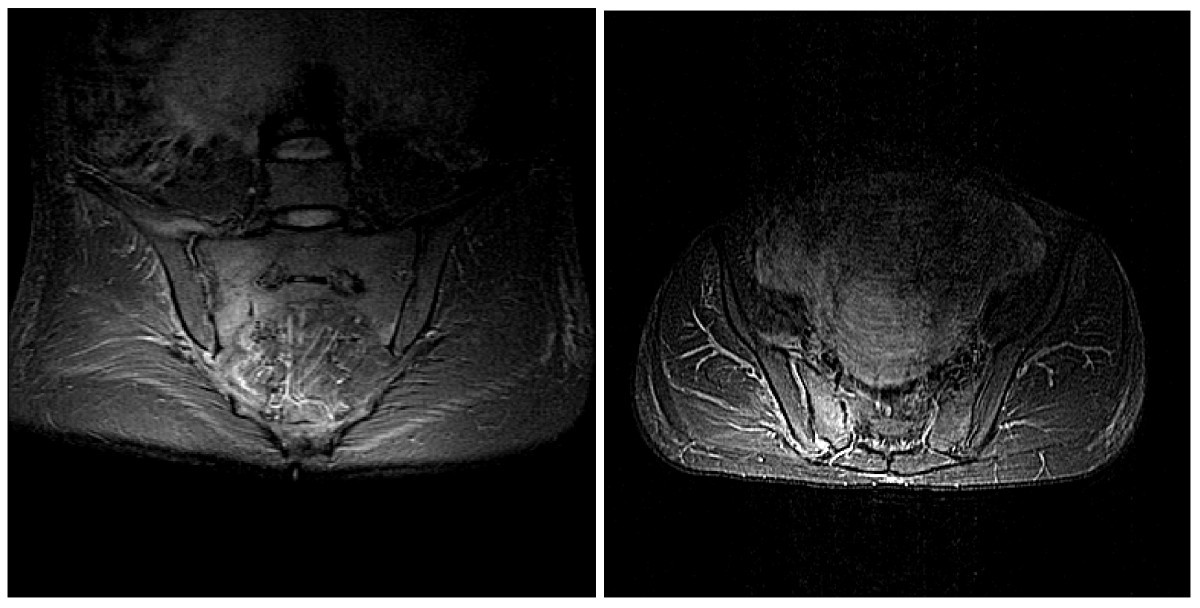
Sacral Spine (S1 - S5) Injuries, Sacral Sparing
13 hours ago · Overall, Positive Predictive Value (PPV) ranged between 48% (VAC) to 73% (DAP) and Negative Predictive Value (NPV) between 92% (VAC) to 100% (LT). AIS-A had NPV of 100% across all categories, and AIS-D had PPV of 100% across all categories. Conclusion: Patient report of sacral sparing can predict negative sensation in patients with AIS-A and predict … >> Go To The Portal
Patient report of sacral sparing can predict negative sensation in patients with AIS-A and predict positive sensation in persons with AIS-D. Overall, the self-report of sacral sparing of motor and sensory function is not predictive enough to rely on for accurate classification. Keywords: Rectal examination; Sacral; Spinal cord injury.
Full Answer
Is sacral examination in spinal cord injury really needed?
Sacral examination in spinal cord injury: Is it really needed? Patient report of sacral sparing can predict negative sensation in patients with AIS-A and predict positive sensation in persons with AIS-D. Overall, the self-report of sacral sparing of motor and sensory function is not predictive enough to rely on for accurate classification.
What are the effects of a sacral spinal cord injury?
Injuries to the sacral spine may leave the patient with some degree of function loss in the hips and/or legs. The patient will most likely be able to walk, and drive a car. An injury to the sacral spinal cord may leave the patient with little or no bladder or bowel control, however, the patient will be completely...
How common are sacral spine injuries?
Injuries to the sacral spine are less common than injuries to other areas of the spine. It is also the least likely area for spinal nerves to compress. The sacrum is the triangle-shaped bone at the end of the spine between the lumbar spine and the tailbone.
Why are sacral reflexes important in the evaluation of spinal cord segments?
In conclusion, sacral reflexes and determination of voluntarily anal sphincter contraction are important to allow the practitioner to gain information about the state of the sacral spinal cord segments. When present, it is indicative of intact spinal reflex arcs and reflex conal autonomic functions.

What are the symptoms of sacral nerve damage?
Damage to the spine at the sacrum levels affects the nerve roots as follows: S1 affects the hips and groin area. S2 affects the back of the thighs....SymptomsLack of control of bowels or bladder.Lower back pain.Leg pain, which may radiate down the back of the leg(s)Sensory issues in the groin and buttocks area.
What is the sacral in the spine?
The sacral spine (sacrum) is located below the lumbar spine and above the tailbone, which is known as the coccyx. Five bones that are fused together make up the triangle-shaped sacrum, and these bones are numbered S-1 to S-5. Each number corresponds with the nerves in that section of the spinal cord.
What is sacral nerve damage?
Patients with sacral nerve injuries may have symptoms on one or both sides of the body. Injuries to the sacral spine may leave the patient with some degree of function loss in the hips and/or legs. The patient will most likely be able to walk and drive a car.
How do you check for sacral sparing?
To determine if a spinal cord injury patient has sacral sparing, an anal exam is required....Your doctor will examine functions innervated by the bottom-most spinal cord segments (S4-S5) like:light touch at the perianal area.pinprick sensation at the perianal area.deep anal pressure.voluntary anal contraction.
What is sacral back pain?
Sacroiliitis (say-kroe-il-e-I-tis) is an inflammation of one or both of your sacroiliac joints — situated where your lower spine and pelvis connect. Sacroiliitis can cause pain in your buttocks or lower back, and can extend down one or both legs. Prolonged standing or stair climbing can worsen the pain.
What is a sacral?
The sacral region (sacrum) is at the bottom of the spine and lies between the fifth segment of the lumbar spine (L5) and the coccyx (tailbone). The sacrum is a triangular-shaped bone and consists of five segments (S1-S5) that are fused together.
What are the 5 sacral nerves?
The sacral plexus is derived from the anterior rami of spinal nerves L4, L5, S1, S2, S3, and S4. Each of these anterior rami gives rise to anterior and posterior branches.
What is sacral area?
The sacrum, sometimes called the sacral vertebra or sacral spine (S1), is a large, flat triangular shaped bone nested between the hip bones and positioned below the last lumbar vertebra (L5). The coccyx, commonly known as the tailbone, is below the sacrum.
How long does sacrum take to heal?
Fracture healing A sacral fracture takes 8–12 weeks to heal and fusion rates following sacral fractures have been reported to be 85–90%. Malunion can occur after delayed treatment or insufficient reduction, with a consequent alteration of pelvic incidence.
Where is the sacral nerve located?
Location. The spinal nerves that comprise the sacral plexus emerge from the lateral (side) regions of the spinal cord. Each of these nerves travels through its corresponding spinal foramen (opening) before they join in their various combinations to form the sacral plexus in the back of the pelvis.
What is the significance of sacral sparing?
Sacral sparing, determines whether a spinal cord injury (SCI) is classified as neurologically complete or incomplete.
What nerve is between L5 and S1?
The lumbar nerve roots exit beneath the corresponding vertebral pedicle through the respective foramen. For example, the L5 nerve root exits beneath the L5 vertebral pedicle through the L5/S1 foramen.
What is the function of sacral vertebrae?
The function of the sacral vertebrae is to secure the pelvic girdle, the basin-like bone structure connect ing the truck and the legs, supporting and balancing the trunk, and containing the intestines, bladder, bowel, and internal sex organs.
Where are the sacral vertebrae located?
The sacral vertebrae are represented by segments S1 through S5 and located between the lumbar vertebrae and the coccyx (tailbone) —the lowest part of the vertebral column.
What is the sacral canal?
The sacral canal runs down the center of the sacrum, representing the end of the vertebral canal. The five segments of the sacral vertebrae affect nerve communication to the lower part of the body. There, numerical levels are often mentioned in imaging studies of the spine. S1 refers to the first sacral bone, S2 to the second sacral bone, and so on.
What is the name of the wing of the sacrum?
The first three vertebrae of the sacral region form the wide lateral wings called the alae. The alae (also called the ala or wing of sacrum) connect with the blades of pelvis—called the ilium . The sacrum also forms the back wall of the pelvis and the joints at ...
What causes sacral vertebrae to be damaged?
Common causes of injuries related to the sacral vertebrae include car accidents, sports injuries, trauma, falls, birth defects, osteoporosis, and joint degeneration . Injuries and damage to S1, S2, S3, S4, or S5 can still leave a person functional, but they primarily affect bowel and bladder function.
How many bones are in the sacral vertebrae?
Treatment. The sacral vertebrae—also called the sacral spine—consists of five sacral vertebrae bones. These bones fuse together to form the sacrum, the shield-shaped bony structure located at the base of the lumbar vertebrae (the five cylindrical bones forming the spine of the lower bank) and connected to the pelvis.
What is the sacrum?
The sacrum also form s the back wall of the pelvis and the joints at the hip bones called the sacroiliac joints . There are a series of four openings on each side of the sacrum where the sacral nerves and blood vessels run. The sacral canal runs down the center of the sacrum, representing the end of the vertebral canal.
What is the sacral region?
The sacral region is composed of five segments, S1 to S5, that are fused together. The sacrum is part of the pelvic girdle and contributes to the formation of joints at the hip bone called the sacroiliac joints. The sacral region contains a serious of four openings on each side through which the sacral nerves and blood vessels run.
How to treat sacrum pain?
Luckily, treatment for sacrum pain usually does not require surgery, as getting adequate rest, taking pain relieving medication, and staying active is often enough to fully resolve the pain over time. Your doctor may recommend you wear a medical brace or corset to help support the bone structure, but this is seldom needed. Water exercises may help maintain flexibility while limiting tension on the back muscles. In severe cases where a fracture has occurred, a sacroplasty procedure may be required, where bonding material is injected into the joint site for faster fusion of the fracture.
What muscles are involved in sacrum pain?
Other muscles that result in pain to this area include the deep lateral rotators, hamstrings, hip flexors, and pelvic floor muscles. A vertical fracture of the pelvic area may also result in sacrum pain. This fracture typically runs parallel to your spine. However, fractures going horizontally across the sacrum are also a possible cause ...
What is the lower back?
The lower back has a diverse set of muscles involved in postural stability and other flexor and extension actions. This makes lower back pain or sacrum pain a complicated diagnosis, as many variables are at play. Various connecting muscles may refer the pain away from the actual site of injury, requiring medical professionals to examine all ...
Why does my sacrum hurt?
What causes pain in the sacrum? Pain in the area of the sacrum can be due to the ligaments becoming too loose or too tight. This may be caused by a fall injury, work injury, car accident, pregnancy, or hip/spine surgery (laminectomy, lumbar fusion). Many diseases may also lead one to experience pain in this region.
What is the best treatment for sacrum pain?
If sacrum pain is due to bone weakness, vitamin D and calcium supplementation may be appropriate.
What is the sacrum?
The sacrum is the key stone of the pelvis and serves several important functions in the skeletal, muscular, nervous, and female reproductive systems. Also, several key muscles such as the gluteus maximus, illacus, and piriformis require their sacrum to aim in moving the leg.
Why are sacral reflexes important?
Sacral reflexes are important to allow the SCI practitioner to gain information about the state of the sacral spinal cord segments. The presence of the bulbocavernosus and/or the anal wink reflex indicate an intact spinal reflex arc and reflex conal autonomic function (as part of the upper motor neuron syndrome);
What is the classification of spinal cord injury?
The International Standards for Neurological Classification of Spinal Cord Injury (ISNCSCI) were developed in 1982 by the American Spinal Injury Association (ASIA) to provide precision in the definition of neurological levels and extent of spinal cord injury (SCI) and achieve consistent and reliable data for patient care and research [ 1 ]. The ISNCSCI gives an accurate neurological level of injury (NLI) and is useful to prognosticate motor recovery [ 2 ]. The International Standards for the Assessment of Autonomic Function after SCI (ISAFSCI) were published in 2009 and designed to complement the ISNCSCI by documenting the impact of SCI on autonomic neural control of specific organ systems, including the sacral responses of bladder, bowel, and sexual function [ 3 ].

Injuries to The Sacral Spine
Causes
- The most common causes of spinal cord injuries to the sacrum are: 1. Motor vehicle accidents 2. Trauma 3. Falls 4. Birth defects 5. Degeneration 6. Osteoporosis
Treatment
- Current treatments available for spinal cord patients with sacrum injuries are: 1. Drugs:Non-steroidal anti-inflammatory (NSAID) drugs are used in treating spinal cord and nerve root injuries. The quicker these drugs are initiated after injury, the better the result for the patient by reducing inflammation around the spinal cord. 2. Surgery:Surgical decompression of the nerves and fusio…
Additional Information
- Damaging either the S1, S2, S3, S4, or S5 vertebrae should leave the patient fairly functional with some issues controlling bowel and bladder function. Patients with injuries to the sacrum typically live very normal lives. Some assistance may be needed for these patients, but most do well on their own.