Paradigm shift: the Copernican revolution in diverticular …
7 hours ago EAES/SAGES consensus conference on acute diverticulitis: a paradigm shift in the management of acute diverticulitis Surg Endosc. 2019 Sep;33(9):2724-2725. doi: 10.1007/s00464-019 … >> Go To The Portal
What is the typical follow-up time for diverticulitis?
Median follow-up time was 537 days (Interquartile Range (IQR): 229–1042 days). Of all patients, 3,844 (32%) had subsequent treatment encounters for recurrent diverticulitis (50% outpatient recurrences, 4% recurrences requiring emergency operation).
How is left lower quadrant pain evaluated in suspected diverticulitis?
The preferred examination for evaluation of acute left lower quadrant pain and suspected diverticulitis is CT of the abdomen and pelvis with oral, rectal, and intravenous (IV) contrast. Acquisition of thin (1 mm or less) axial source images allows generation of high-quality reconstructed 3 mm axial and coronal images.
Does surgery improve quality of life in patients with diverticulitis?
A recent trial comparing elective surgery and conservative management for patients with recurrent diverticulitis revealed that surgery was associated with significantly higher GI-specific quality of life (DIRECT trial),10emphasizing the importance of patient-specific outcomes in this disease.
What is the pathophysiology of diverticulitis?
Diverticulitis- Stool passing slowly through the intestine deposits fecal material in the diverticula. Over time, bacterial overgrowth causes an inflammatory response and may form an abscess or infection in the diverticula.

What is the gold standard for assessing diverticulitis?
Multidetector CT is now considered the gold standard for assessing this disease. The preferred examination for evaluation of acute left lower quadrant pain and suspected diverticulitis is CT of the abdomen and pelvis with oral, rectal, and intravenous (IV) contrast.
What is CT for diverticulitis?
CT is helpful in identifying and/or excluding other causes of abdominal pain when diverticulitis is not the etiology. Differential diagnosis includes neoplasm, appendicitis, epiploic appendagitis, ischemic colitis, and inflammatory bowel disease.
Is diverticulosis asymptomatic or asymptomatic?
Because the incidence of colonic diverticulosis is high in the general population, incidental asymptomatic diverticulosis is commonly seen on radiology imaging studies. However, diagnostic imaging performed specifically for diverticular disease is essentially limited to imaging of suspected acute colonic diverticulitis (ACD) and its complications.
Is plain film radiography good for diverticulitis?
Plain film radiography is usually of little value in the assessment of suspected diverticulitis unless there is free intraperitoneal air from perforation, portal venous gas, or signs of bowel ileus or obstruction. However, these findings are nonspecific.
Is diverticulosis a mitigating factor?
The presence of diverticula in an involved segment suggests diverticulitis, but the high prevalence of diverticulosis in the general population dictates that the presence of diverticula cannot be used as a mitigating factor to exclude neoplasm.
Can small bowel pathology mimic diverticulitis?
Small bowel pathology can also mimic diverticulitis, and CT is helpful to distinguish these patients. Epiploic appendagitis is a nonsurgical entity that results from torsion and venous occlusion of an epiploic appendage, and patients present with signs and symptoms that mimic diverticulitis.
Is diverticulitis a neoplasm?
Colonic wall thickening is usually greater (measuring >2 cm) with neoplasm and is more often eccentric. Diverticulitis and neoplasm may both involve short segments of bowel; however, when a segment of >10 cm is involved, this is considered specific for diverticulitis.
Where are diverticula found?
These pouches may form anywhere along the intestine, but are most commonly found at the end of the descending and sigmoid colons on the left side of the abdomen. They are also commonly found in the first section of the small intestine, but diverticula in this area rarely cause problems. Diverticulitis: involves small abscesses or infection in one ...
What is the term for stool passing slowly through the intestine deposits fecal material in the diverticul
Diverticulitis- Stool passing slowly through the intestine deposits fecal material in the diverticula. Over time, bacterial overgrowth causes an inflammatory response and may form an abscess or infection in the diverticula.
How to prevent constipation?
Maintain optimal hydration for improved intestinal motility to prevent constipation. Administer medications. Antibiotics – for infection. Analgesics – for pain. IV Fluids – for hydration and bowel motility. Psyllium – (bulk-forming laxative) absorbs water from the intestine and makes stool easier to pass.
What is the term for a stool that is hard and slow to pass through the intestine?
These pockets are most often asymptomatic. Diverticulitis- Stool passing slowly through the intestine deposits fecal material in the diverticula.
Where are diverticulosis pouches found?
These pouches may form anywhere along the intestine, but are most commonly found at the end of the descending and sigmoid colons on the left side of the abdomen. They are also commonly found in the first section of the small intestine, but diverticula in this area rarely cause problems. Diverticulitis: involves small abscesses or infection in one or more of the diverticula, or perforation of the bowel.
What is the best way to get rid of constipation?
Psyllium – (bulk-forming laxative) absorbs water from the intestine and makes stool easier to pass. Provide nutrition education. Hydrate (2-3 L fluids daily, unless contraindicated for renal or cardiac disease) to avoid constipation. Probiotics – to help regulate the intestinal bacteria.
Can diverticulosis cause pain?
A patient that has diverticulosis without the diverticulitis may not experience any pain at all. In fact, they may not even realize they have the disease. At first, the patient that starts to get that diverticulitis, where those diverticula, the intestines become inflamed and irritated and infected.
How to prevent diverticulitis?
Preventing Recurrence. Reach for Foods High in Fiber. Fiber is a form of carbohydrate that helps keep the digestive system healthy. Studies suggest that eating enough fiber can help you avoid recurrence of diverticulitis and reduce the need for surgery in the future.
Can diverticulitis be treated without surgery?
Acute diverticulitis is a painful, relatively sudden condition that can usually be treated without surgery. Approximately 20% of patients with diverticulitis will have another flare-up in the future. There may be several steps you can take to lower the risk of future attacks.
Can diverticulitis cause colon cancer?
Talk to Your Doctor About Getting a Colonoscopy After Your Condition Improves. In rare cases, diverticulitis can actually be a warning sign for colon cancer.
Introduction
Diverticula are outpouchings of the intestinal wall lining. When these pouches become inflamed or infected, they can lead to diverticulosis (inflammation) or diverticular disease (infection). These conditions can cause significant pain in your abdomen and other signs and symptoms like fever or nausea.
What is Diverticulitis?
Diverticulitis is an inflammation of the intestine that most often occurs in the colon. The inflammation is caused by a bacterial infection of any one or more of the pouches created as part of diverticulosis. The inflammation can come on suddenly, or you may have a history of gradual onset over 24 to 48 hours.
What Causes Diverticulitis?
Diverticulitis may be caused by poor diet intake (such as low dietary fiber or too much fat), obesity, and age. The weakening of the walls can be attributed to long bouts of constipation.
Nursing Diagnosis of Diverticulitis
Your doctor will feel for tenderness, swelling, and possibly a mass or lump in your colon. The presence of either means that diverticulitis has developed.
Risk Factors for Diverticulitis
1) Diverticular Abscess (acute): Risk for impaired gas exchange secondary to respiratory infection or pulmonary edema.
Nursing Care Plan for Diverticulitis
The patient should have a high fiber diet to help prevent constipation. Give clear fluids only if tolerated.
Nursing Interventions for Diverticular Abscess (Acute)
Monitor temperature and pulse, watch for respiratory status changes. Ask the patient about symptoms of pain, nausea, vomiting to determine the level of distress. Offer sips of water every 1-2 hours to prevent dehydration. Assess the amount, color and character (thick or thin), urine output.
What is complicated diverticulitis?
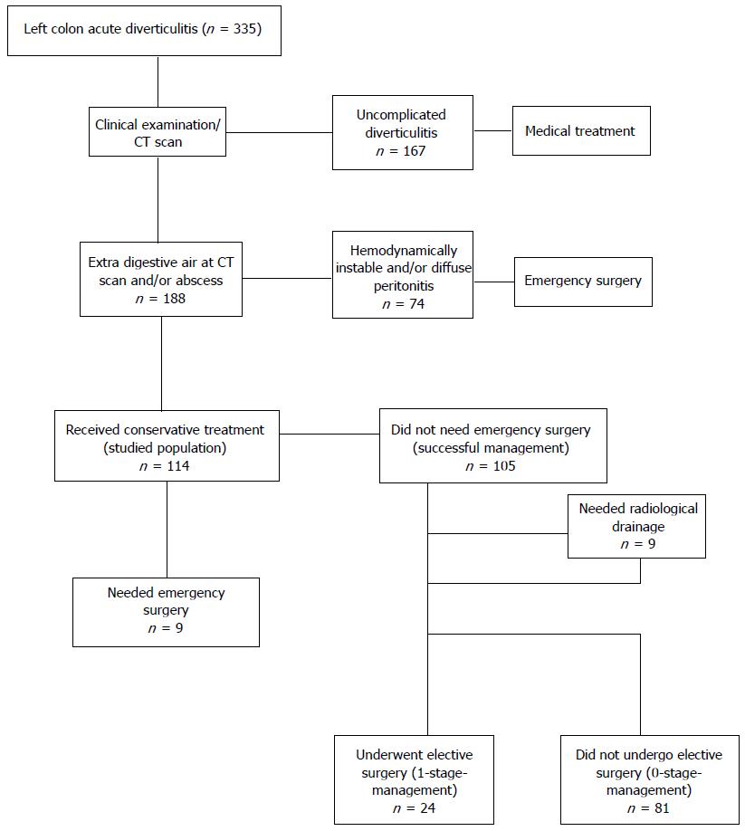
complicated diverticulitis is defined as diverticulitis with 1 of the following associated complications. bowel obstruction. abscess. fistula. perforation. Epidemiology. demographics. most commonly occurs at the sigmoid colon in North America, reflecting the distribution of diverticulosis.
How long do you need to take antibiotics for diverticulitis?
oral antibiotics for 7-10 days with following 2-3 days after first visit. Inpatient medical management.
What is the temperature of a 70 year old man with chronic constipation?
He is found to have a temperature of 100.8F, BP 140/90, HR 85, and RR 16. On physical examination, he is tender to light palpation in the left lower quadrant and exhibits voluntary guarding. Rectal examination reveals heme-positive stool. Laboratory values are unremarkable except for a WBC count of 12,500 with a left shift. Which of the following tests would be most useful in the diagnosis of this patient's disease?
What are the positive findings of abdominal ultrasound?
indicated in patients who cannot receive radiation. positive findings include bowel wall thickening, hypoechoic peridiverticular inflammatory reaction, and the presence of diverticula. abdominal and chest radiographs.

Popular Posts:
- 1. heywood healthcare patient portal
- 2. washakie patient portal
- 3. miford regional patient portal
- 4. summithealth patient portal
- 5. foothills neurology patient portal
- 6. tmh craig patient portal
- 7. pulmcare.com patient portal
- 8. university of mo. hospital patient portal
- 9. family healthcare partners patient portal grove city pa
- 10. tri county eye patient portal