What Is Optical Coherence Tomography? - American …
22 hours ago · Optical coherence tomography is a quick, non invasive and reproducible imaging tool for macular lesions and has become an essential part of retina practice. This review address the common protocols for imaging the macula, basics of image interpretation, features of common macular disorders with clues to differentiate mimickers and an introduction to … >> Go To The Portal
The optical coherence tomography confirmed the reduction of foveal depression on the first case and the disarrangement of all retinal layers on the second. There has been complete functional and anatomical resolution by optical coherence tomography in the first patient, while the second evolved to permanent visual loss.
Full Answer
What is Oct exam eye?
The ability to obtain in vivo images of the vitreous has greatly advanced in the optical coherence tomography (OCT) era. Swept-source OCT (SS-OCT) was especially helpful because it has less roll-off of the signal with increasing depth in the tissue.
How to read macular Oct?
References
- Huang D, Swanson EA, Lin CP, Schuman JS, Stinson WG, Chang W, et al. Optical coherence tomography. ...
- Diabetic Retinopathy Clinical Research Network Writing Committee. Bressler SB, Edwards AR, Chalam KV, Bressler NM, Glassman AR, et al. ...
- Diabetic Retinopathy Clinical Research Network. ...
What is an Oct eye exam?
Several eye conditions can be diagnosed using OCT, including:
- Glaucoma
- Age-related macular degeneration (AMD)
- Diabetic retinopathy
- Central serous retinopathy
- Macular hole
- Macular edema
- Macular pucker
- Vitreous traction
- Optic nerve disorders
What does tomography, optical mean?
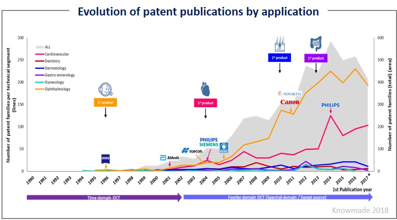
Optical Coherence Tomography (OCT) is a technique for obtaining sub-surface images such as diseased tissue just below the skin. Ophthalmologists use OCT to obtain detailed images from within the retina. Cardiologists also use it to help diagnose coronary artery disease.

How do you describe an OCT patient?
OCT explanations When discussing OCT with the patient, you must make a spectacle of it; it's all about a little bit of theatre. We have found that using 3D scans of the eye is a good way to show off OCT to patients and illustrate to them how the technology works. We do so in the test room on 55 inch monitors.
How do I read my OCT RNFL report?
8:4317:21Then the machine check an area four millimeters centered around the desk. As you can see this colorMoreThen the machine check an area four millimeters centered around the desk. As you can see this color part you get a clue here of the thickness of the inner fiber layer.
How do you read OCT findings?
Interpretation of the OCT image requires understanding that the image was based on light waves. Thus, the image is subject to the characteristics of light waves, and one can see reflections, attenuations, interfaces, and shadows in the image due to this property of light.
What does an OCT scan of the eye show?
OCT provides detailed images of the retina and macula, showing irregularities caused by AMD. With OCT imaging, your optometrist can detect signs of AMD like drusen (tiny clumps of protein) caused by dry AMD and abnormal blood vessels and bleeding caused by wet AMD.
What is normal RNFL thickness?
Average RNFL thickness indicates a patient's overall RNFL health. The mean value for RNFL thickness in the general population is 92.9 +/- 9.4 microns. Typically, a normal, nonglaucomatous eye has an RNFL thickness of 80 microns or greater.
What is normal RNFL symmetry?
Mean RNFL thickness between the 2 eyes of normal individuals should not differ by more than approximately 9 to 12 μm, depending on which scanning algorithm of OCT3 is used and which eye measures thicker.
What is normal macular thickness on OCT?
The OCT software determined the center (mean ± SD central foveal thickness) to be 207 ± 18 μm.
Can OCT scan detect glaucoma?
Optical coherence tomography is particularly effective at detecting early and preperimetric glaucoma in the office setting. Its use for screening works best when the technology is combined with a clinical eye examination or with other strategies such as portable tonometry, fundus photography, and visual field testing.
Does OCT show retinal detachment?
OCT can detect signs of glaucoma, retinal detachment, macular degeneration, diabetic retinopathy and more at an incredibly early stage.
Can an OCT scan detect cataracts?
In individual patients, OCT scans remain reliable for gross clinical interpretation, even in the presence of cataract.
Can an OCT scan detect diabetes?
When the blood vessels in the central area of the retina (the macula) are affected, it's known as diabetic maculopathy. When these blood vessels are damaged they become leaky and cause swelling of the back of the eye which can be detected easily by an OCT scan.
Can an OCT detect brain tumor?
The researchers are using OCT imaging to determine the tumor boundaries.
Abstract
Optical coherence tomography is a quick, non invasive and reproducible imaging tool for macular lesions and has become an essential part of retina practice.
Optical Coherence Tomography Scan Protocols
Scan protocols used in the more widely used SD-OCT systems are mentioned in this section. The commonly used scan protocols for macular scanning are three-dimensional (3D) scan, radial scan, and raster scan [ Fig. 3 ]. A 3D scan consists of a number of horizontal line scans [ Fig.
Optical Coherence Tomography Scan Acquisition Procedure
A minimum pupil diameter of 3 mm is required to obtain a good OCT image. An appropriate scanning protocol is selected to scan the retinal area of interest and a live OCT window is seen. The patient is instructed to look at the internal target at the center (for macular scanning) or an external target where appropriate.
Measurements
Retinal thickness is a reproducible and common quantitative measurement that is used to monitor the disease process or treatment response using OCT.
Artifacts on Optical Coherence Tomography
OCT artifacts could be patient related, operator-related, and software related. While patient- and operator-related artifacts can be controlled to some extent, software-related errors are inevitable and most common. Patient-related artifacts are mostly due to eye movements, which can be controlled by eye tracking software.
Optical Coherence Tomography Features in Common Macular Disorders
Being noninvasive, quick and reproducible, OCT is used commonly in the diagnosis, and the management of optic nerve and retinal disorders not only for diagnosis but also as a follow-up tool both in clinical practice and in many multicentric trials.
Optical Coherence Tomography in Disorders of the Vitreomacular Interface
These include vitreomacular adhesion (VMA), vitreomacular traction (VMT), and macular hole.
What is OCT used for?
In the setting of cardiology, OCT is used to image coronary arteries in order to visualize vessel wall lumen morphology and microstructure at a resolution 10 times higher than other existing modalities such as intravascular ultrasounds, and x-ray angiography ( intraco ronary optical coherence tomography ). For this type of application, approximately 1 mm in diameter fiber-optics catheters are used to access artery lumen through semi-invasive interventions such as percutaneous coronary interventions .
What is optical coherence tomography?
Optical coherence tomography is based on low-coherence interferometry, typically employing near-infrared light. The use of relatively long wavelength light allows it to penetrate into the scattering medium.
What is OCT imaging?
Optical coherence tomography, (OCT), is a technique for obtaining sub-surface images of translucent or opaque materials at a resolution equivalent to a low-power microscope. It is effectively ‘optical ultrasound’, imaging reflections from within tissue to provide cross-sectional images.
How does spatially encoded frequency domain OCT work?
Spatially encoded frequency domain OCT (SEFD-OCT, spectral domain or Fourier domain OCT) extracts spectral information by distributing different optical frequencies onto a detector stripe (line-array CCD or CMOS) via a dispersive element (see Fig. 4). Thereby the information of the full depth scan can be acquired within a single exposure. However, the large signal to noise advantage of FD-OCT is reduced due to the lower dynamic range of stripe detectors with respect to single photosensitive diodes, resulting in an SNR ( signal to noise ratio) advantage of ~10 dB at much higher speeds. This is not much of a problem when working at 1300 nm, however, since dynamic range is not a serious problem at this wavelength range.
Why is OCT so high resolution?
No ionizing radiation. OCT delivers high resolution because it is based on light, rather than sound or radio frequency. An optical beam is directed at the tissue, and a small portion of this light that reflects from sub-surface features is collected.
What are the benefits of OCT?
The key benefits of OCT are: 1 Live sub-surface images at near-microscopic resolution 2 Instant, direct imaging of tissue morphology 3 No preparation of the sample or subject, no contact 4 No ionizing radiation
What is FD OCT?
In frequency domain OCT (FD-OCT) the broadband interference is acquired with spectrally separated detectors. Two common approaches are swept-source and spectral-domain OCT. A swept source OCT encodes the optical frequency in time with a spectrally scanning source. A spectral domain OCT uses a dispersive detector, like a grating and a linear detector array, to separate the different wavelengths. Due to the Fourier relation ( Wiener-Khintchine theorem between the auto correlation and the spectral power density) the depth scan can be immediately calculated by a Fourier-transform from the acquired spectra, without movement of the reference arm. This feature improves imaging speed dramatically, while the reduced losses during a single scan improve the signal to noise ratio proportional to the number of detection elements. The parallel detection at multiple wavelength ranges limits the scanning range, while the full spectral bandwidth sets the axial resolution.
What eye conditions can be diagnosed with OCT?
Macular edema. Macular pucker. Vitreous traction. Optic nerve disorders. Not only can OCT help diagnose eye conditions, it can also monitor certain problems that are already present. Glaucoma is one such condition that can be monitored with OCT, as the exam helps ophthalmologists detect changes that occur in the optic nerve fibers.
Why is OCT important?
This imaging is important for detecting and monitoring changes in the retina and optic nerve. OCT can help detect a number of retinal diseases, such as glaucoma, diabetic retinopathy and age-related macular degeneration (AMD). Additionally, OCT can be used to monitor some neurological conditions such as multiple sclerosis (MS).
What is OCT scan?
OCTA is derived from OCT technology. An OCTA scan provides just as much information as OCT, but with more advanced, three-dimensional images. The imaging is particularly useful in detecting and treating inflammatory conditions such as uveitis because of the technology.
What is OCTa in the eye?
OCTA also provides insight into the blood flow within both the retina and the choroid (the layer of tissue between the sclera [white of the eye] and retina). Although OCTA provides great detail for eye doctors (and their patients), there are still some limitations due to its newness.
Why is OCT not useful?
Although OCT can be used for many different eye conditions, it is not useful for those that affect the way light passes through the eye , such as cataracts. This is because OCT technology depends upon light waves for proper exam readings and results.
Can OCT be used for MS?
Although MS is usually monitored with magnetic resonance imaging (MRI), clinicians have found that OCT technology can provide additional imaging of the retinal nerves in MS patients and provide a greater assessment of the disease.
Does myelin protect nerve fibers?
Myelin also does not protect retinal nerve fibers, and research has shown that these fibers actually thin out in patients with MS. Because of this lack of protection, clinicians are able to get a better look at isolated axons during OCT exams (and evaluate the retinal nerve fibers at the same time).

Popular Posts:
- 1. wcmc patient portal
- 2. alpharetta internal medicine patient portal
- 3. wright state physicians patient portal login
- 4. memorial hospital patient portal ri
- 5. patient portal partners in health
- 6. dr bruce davis family practice patient portal
- 7. how to sign up for shriners patient portal
- 8. patient portal chesapeake urology
- 9. https:www.covenantmedicalgroup.org/secure-patient-portal
- 10. adventhealth wesley chapel patient portal