How to Read Your Radiology Report
23 hours ago · Next, the radiologist writes a report detailing the results. A typical radiology reports includes these sections: Name or Type of Exam; Date of Exam; Interpreting Radiologist – the name of the radiologist who read the diagnostic imaging exam and wrote the report. Clinical History – … >> Go To The Portal
How does the patient access the radiologist’s report?
The patient accesses the radiologist’s report through a secure portal on the Internet at the same time as the referring physician or soon after. At many hospitals and imaging facilities, especially academic centers, this shift in practice already has begun.
Can I Read my radiology reports online?
Many patients can read their electronic health records online. Sometimes, these records include radiology reports. Online access to your health records may help you make more informed decisions about your healthcare. In addition, online access lets you share your radiology reports with other doctors electronically.
Can an MRI scan diagnose MS by itself?
MRI scans are an important way to help health care providers figure out if a person has MS or not, but MRI scans cannot diagnose MS by themselves. While it is true that almost all people with MS will have lesions on MRI, not all people with MRI lesions have MS.
How do I review my MRI report?
Reviewing an MRI report is best accomplished face-to-face or over the phone with a health care provider who can explain the language of the report, the significance of any MRI findings, and interpret the report in the broader context of your overall condition.

How do you read multiple sclerosis MRI?
0:406:58Multiple Sclerosis: Understanding Your MRI - YouTubeYouTubeStart of suggested clipEnd of suggested clipThe areas of loss of myelin. Leave their marks seen on MRI that appear as spots called lesions. MostMoreThe areas of loss of myelin. Leave their marks seen on MRI that appear as spots called lesions. Most new activities seen on MRI.
How do I read my MRI results?
MRI interpretation Systematic approachStart by checking the patient and image details.Look at all the available image planes.Compare the fat-sensitive with the water-sensitive images looking for abnormal signal.Correlate the MRI appearances with available previous imaging.Relate your findings to the clinical question.
Can MS be seen on xray?
Your doctor can do a rapid test to determine if you have strep throat, and see your broken arm using an X-ray. Unfortunately, multiple sclerosis (MS) is much more complex and can take months, or even longer, to accurately diagnosis. There is no single test that will tell you if you have MS.
What does an MRI report look like for MS?
MS activity appears on an MRI scan as either bright or dark spots. Typical MS lesions tend to be oval or frame shaped. MS lesions can appear in both the brain's white and gray matter. Healthcare professionals may use a chemical contrast dye called gadolinium to improve the brightness of MRI scan images.
What is radiologist report?
A radiology report is the official record of medical images that contains the interpretations and images [1]. The main goal of the radiology report is to present the outcomes of the imaging procedure (e.g. X-ray, MRI) of the patients to physicians [2].
What makes a good radiology report?
The report should communicate relevant information about diagnosis, condition, response to therapy, and/or results of a procedure performed. The written report should also answer any clinical question raised by the requesting patient-care provider that is relevant to the radiologic study.
How many lesions is alot for MS?
According to the team, patients with a combination of more than 13 lesions, with a maximal lesion diameter greater than 0.75 cm, and lesions perpendicular to the corpus callosum, had a 19 times greater chance of progressing to MS during the following year.
Does white matter on brain mean MS?
White Matter Consists Mainly of Nerve Fibers In MS, the immune system attacks the myelin in the brain, spinal cord, and optic nerves. The attack causes inflammation that eventually leads to sclerosis, which is the medical term for scarring. (That's how MS got its name.)
How do I know if my MS is progressing?
To figure out if disease is progressing, doctors use a scale called the Expanded Disability Status Scale (EDSS). The EDSS is a way of measuring physical disability. Two-thirds of those with MS will not progress past level 6 on the EDSS.
What are T2 lesions in MS?
T2 weighted imaging identifies MS lesions as high signal foci against the low signal background of white matter. However, periventricular lesions are often indistinguishable from the adjacent CSF which is also of high signal with T2 weighting.
What does T1 and T2 mean on MRI?
The most common MRI sequences are T1-weighted and T2-weighted scans. T1-weighted images are produced by using short TE and TR times. The contrast and brightness of the image are predominately determined by T1 properties of tissue. Conversely, T2-weighted images are produced by using longer TE and TR times.
What is increased T2 signal on MRI report brain?
A hyperintensity or T2 hyperintensity is an area of high intensity on types of magnetic resonance imaging (MRI) scans of the brain of a human or of another mammal that reflect lesions produced largely by demyelination and axonal loss.
What does "impression" mean in a radiologist?
Impression – this is the radiologist’s “impression” or diagnosis of the diagnostic imaging exam. This section includes a summary of the results and any follow up testing (like a biopsy or additional diagnostic imaging) that the radiologist recommends. ...
What is included in a radiology report?
A typical radiology reports includes these sections: Name or Type of Exam. Date of Exam. Interpreting Radiologist – the name of the radiologist who read the diagnostic imaging exam and wrote the report. Clinical History – describes the patient’s symptoms or existing diagnosis.
Why do radiologists need to talk to patients about their history?
Conversations with patients about their clinical histories can ensure that physicians have access to all relevant data before referring the patient for diagnostic imaging.
Why do radiologists use web portals?
Increasingly, diagnostic imaging providers use web portals to give patients full access to their own radiology reports. Despite early fears that patients would misinterpret complex medical information, leading to unnecessary anxiety, most studies of direct access to radiology reports suggest that this is a positive step for patients and health care providers alike. When patients take a more active role in their care, they provide an extra level of oversight, reducing the chance of an error. Informed patients are also more likely to ask radiologists helpful questions, potentially preventing unnecessary testing.
What is a comparison section in a radiology report?
If the patient has had other imaging done in the past, sometimes those images need to be used as comparisons. The “comparison” section of the typical radiology report is where past images will be mentioned. Typically, radiologists only consult scans of similar regions of the body when making comparison notes.
What is section 1 of a radiology report?
Section 1: The Basics. Radiology reports that begin with naming the type of exam will explain which imaging modality (MRI, CT, X-ray, etc.) was used in the procedure — and, crucially, which part of the body technologists scanned. They’ll also include the time and date, as well as any details about preliminary procedures, like the use of contrast.
What is the impression section of a medical report?
The “impression” section of a medical report is where patients will find the radiologist’s diagnosis, along with recommendations for future testing for confirmation. Commonly, radiologists offer differential diagnoses, which include any number of potential causes of the patient’s symptoms.
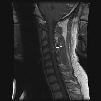
Understanding Your MRI Report
Magnetic resonance imaging, or MRI, is a wonderful tool to help diagnose and follow people with MS. MRI is safe and relatively non-invasive yet can provide very detailed images of the brain and spinal cord that can reveal MS lesions (also known as demyelination, spots, or plaques) and changes in MS activity over time.
Is it MS or not?
MRI scans are an important way to help health care providers figure out if a person has MS or not, but MRI scans cannot diagnose MS by themselves. While it is true that almost all people with MS will have lesions on MRI, not all people with MRI lesions have MS.
Is my MS getting worse?
By itself, an MRI report cannot tell whether or not a person with MS is doing well. Some people have a lot of MS lesions but are doing very well clinically. Some people with just a few MS lesions can be significantly disabled. In general, though, the fewer MS lesions a person has the better.
How long does it take to evaluate a report?
Readers most frequently evaluated reports in less than 2 minutes. When reports and images were evaluated together, no difference in terms of question response was observed for any reader. Reader 1 evaluated reports and MR images in 2–5 minutes; reader 2, in 5–10 minutes; and reader 3, in more than 10 minutes.
How many people are affected by MS?
Multiple sclerosis (MS) is a complex disease of the CNS that is characterized by both inflammatory and neurodegenerative processes and affects about 2.3 million people globally [ 1 ]. CNS MRI is the main tool for diagnosis and monitoring of the disease [ 2 ].
What are the juxtacortical and cortical lesions?
Juxtacortical and cortical lesions are specific for MS. They are adjacent to the cortex and must touch the cortex (yellow circle). In small vessel disease the U-fibers are typically spared and on T2 and FLAIR there will be a dark band of normal WM between the WML and the bright cortex (white circle).
How long does Gadolinium enhancement last?
These enhancing lesions all are new lesions, since Gadolinium enhancement is only visible for about 1 month. So this finding is proof of dissemination in time.
What is a tumefactive MS?
Tumefactive MS is a variant of Multiple Sclerosis.# N#On MRI it presents as a large intra-parenchymal lesion with usually less mass effect than would be expected for its size.#N#They may show some peripheral enhancement, often with an incomplete ring unlike gliomas or intraparenchymal abscesses, which typically have a closed-ring enhancement.
What is the most common inflammatory demyelinating disease of the central nervous system in young and middle-age
Diagnosis of multiple sclerosis: 2017 revisions of the McDonald criteria. Multiple sclerosis is the most common inflammatory demyelinating disease of the central nervous system in young and middle-age adults, but may also affect older people.
What is the McDonald criteria for MS?
According to the McDonald criteria for MS, the diagnosis requires objective evidence of lesions disseminated in time and space, either clinically or radiologically and elimination of more likely diagnoses.
Why is MRI important for MS?
There is an important role for MRI in the diagnosis of MS, since MRI can show multiple lesions - dissemination in space, many of which are clinically occult already at the time of first presentation, and MRI can show new lesions on follow up scans - dissemination in time, much earlier than new symptoms develop. Introduction.
What is the differential between neuromyelitis and myelitis?
A very important differential to keep in mind, especially in patients with a bilateral optic neuritis and myelitis, is Neuromyelitis Optica Spectrum Disorder (NMOSD), previously called Devic's Disease.#N#This is a demyelinating disease caused by antibodies against aquaporin or MOG in which the optic nerves and spinal cord are usually involved.
Why did the PA radiology society oppose the bill?
Berlin says the Pennsylvania Radiological Society, among others, opposed the bill because members feared patients wouldn’t be able to understand the reports. Taxin says there also was opposition because the complex bill was poorly written and would have been costly for radiology practices and hospitals to implement.
What is the piece of the puzzle that a radiology report is?
The radiology report is only one piece of the puzzle, and patients recognize that their doctors have the other pieces, such as medical history, symptoms, and physical exam. Their doctors are the ones who can put all the pieces together to reach a diagnosis and suggest treatment options, he says.
Why are radiologists opposed to providing their reports directly to patients?
One of the naysayers’ biggest concerns was that patients wouldn’t be able to understand the content of the reports and could easily misinterpret the results for the worst.
How long does it take for a radiologist to report a scan?
The radiologist reads the scan and sends the report to the referring physician. The referring physician reports the results to the patient in a few days to a week.
What would happen if you didn't understand the medical reports?
According to Johnson, the patients also said that if they didn’t understand the reports, they would take steps to have them translated into lay terms. Some said they would do their own research on the Internet; some said they would ask friends and family who were more knowledgeable about medical terms.
Can radiologists discuss cancer results?
Taxin says some radiologists probably aren’t comfortable discussing results with patients , especially cancer studies, and never will be. “There are radiologists who just are not used to doing that and won’t get used to it,” he says. “For others, it’s natural.”.
Do radiologists have to send mammography reports?
About three years ago, a Pennsylvania state representative proposed a bill that would require radiologists to send reports of all exams directly to patients. The representative was the friend of a lawyer who was outraged when his wife wasn’t told of her cancer findings and died, Berlin says.
Popular Posts:
- 1. sumner county regional hospital patient portal
- 2. mount nittany medical center patient portal
- 3. forrest general patient portal
- 4. kingwood medical center patient portal
- 5. stony brook psychiatric patient portal
- 6. patient portal technology penn medicine contact number
- 7. lucinda everett patient portal
- 8. ceenta.com patient portal
- 9. patient portal centientall pediatrics
- 10. is there a difference between the doctor's records and patient portal summaries