Brain Imaging in Differential Diagnosis of Dementia
10 hours ago Despite declining ED visits, ED brain imaging in patients with Alzheimer disease and vascular dementia has increased. Various patient-specific and hospital-specific factors contribute to differential utilization rates. Keywords: Alzheimer disease; brain imaging; emergency department; utilization; vascular dementia. >> Go To The Portal
How can brain imaging help professionals with dementia?
Brain Imaging Can Aid Professionals. For instance, vascular dementia may not show evidence of a cortical loss, whereas this qualifier could still allow a patient to have Alzheimer's disease. Also, by undergoing frequent scans, neurologists can make a determination of how the disease is progressing.
What is the future of neuroimaging in dementia management?
It is expected that in future different neuroimaging techniques will increasingly be combined in the management of people with or at risk of dementia with the aim of identifying the presence of a molecular abnormality, studying its effect on the brain structure and function, and measuring the effects of new treatments.
What does a PET scan of the brain show for dementia?
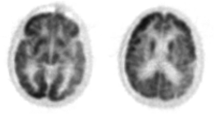
SPECT and PET are similar kinds of scans, and in most cases of degenerative dementia, can showcase bilateral, biparietal, and bitemporal hyperperfusion. Some ligand compounds (when utilized as part of the scan) can reveal the impaired integrity of presynaptic dopamine transporters, present both in degenerative dementias and Parkinson's disease.
How can you tell if someone has dementia in their brain?
Signs Of Dementia In The Brain. Signs accruing and developing inside the brain are more significant, and may help to make a more formal determination of the type of dementia affecting the patient. Brain imaging, such as MRI or PET scans, can reveal these signs and contribute to a more accurate diagnosis.
Does dementia show on a brain scan?
Dementia brain scans Like memory tests, on their own brain scans cannot diagnose dementia, but are used as part of the wider assessment. Not everyone will need a brain scan, particularly if the tests and assessments show that dementia is a likely diagnosis.
What imaging shows dementia?
Computerized tomography (CT) A CT scan is a type of X-ray that uses radiation to produce images of the brain or other parts of the body. A head CT can show shrinkage of brain regions that may occur in dementia, as well as signs of other possible sources of disease, such as an infection or blood clot.
What does MRI look like with dementia?
In the early stages of Alzheimer's disease, an MRI scan of the brain may be normal. In later stages, MRI may show a decrease in the size of different areas of the brain (mainly affecting the temporal and parietal lobes).
Can you see dementia in a brain MRI?
“Can MRI show if I have dementia?” In fact, we scan patients every day with a diagnosis of dementia, memory loss, Alzheimer's, and confusion, among a variety of other neurological disorders. The truth is that MRI is NOT the test to formally diagnose dementia.
What does dementia do to the brain?
Signs and symptoms of dementia result when once-healthy neurons, or nerve cells, in the brain stop working, lose connections with other brain cells, and die. While everyone loses some neurons as they age, people with dementia experience far greater loss.
Which is an early warning signs of dementia?
The 10 warning signs of dementiaSign 1: Memory loss that affects day-to-day abilities. ... Sign 2: Difficulty performing familiar tasks. ... Sign 3: Problems with language. ... Sign 4: Disorientation to time and place. ... Sign 5: Impaired judgement. ... Sign 6: Problems with abstract thinking. ... Sign 7: Misplacing things.More items...
Do white spots on the brain mean dementia?
Conclusion White matter lesions, especially in the periventricular region, increase the risk of dementia in elderly people. Cerebral white matter lesions (WML) in elderly people are thought to result from small-vessel disease and are considered to be a risk factor for dementia.
What do white spots on brain MRI mean?
What Are White Spots? Spots on a brain MRI are caused by changes in water content and fluid movement that occur in brain tissue when the brain cells are inflamed or damaged. These lesions are more easily seen on T2 weighted images, a term that describes the frequency (speed) of the radio impulses used during your scan.
How do you read a brain MRI?
MRI interpretation Systematic approachStart by checking the patient and image details.Look at all the available image planes.Compare the fat-sensitive with the water-sensitive images looking for abnormal signal.Correlate the MRI appearances with available previous imaging.Relate your findings to the clinical question.
What does it mean when an MRI shows white matter?
White matter disease is commonly detected on brain MRI of aging individuals as white matter hyperintensities (WMH), or 'leukoaraiosis.” Over the years it has become increasingly clear that the presence and extent of WMH is a radiographic marker of small cerebral vessel disease and an important predictor of the life- ...
How does a doctor diagnose dementia?
Doctors diagnose the cause of dementia by asking questions about the person's medical history and doing a physical exam, a mental status exam, and lab and imaging tests. Tests can help the doctor find out if the loss of mental abilities is caused by a condition that can be treated.
Are white spots on the brain normal?
Studies have found that white matter lesions appear in some degree on brain scans of most older adults but less often in younger people. White matter lesions are among the most common incidental findings—which means the lesions have no clinical significance—on brain scans of people of any age.
What is brain imaging?
Brain imaging is a way that doctors can make a formal determination of dementia types and progression. Patients exhibit multiple cognitive and behavioral symptoms upon entering the earliest stages of dementia, but these external signs are not the only indications that a physician uses to determine a patient's mental health.
Why do we use scans for dementia?
Using scans to analyze signs of dementia can exclude the possibility of lesions that cause cognitive degeneration or impairment (such as a tumor, an abscess, or a subdural hematoma). It can also help to determine which kind of dementia a patient is experiencing.
What is structural imaging?
Structural imaging of the brain consists of computed tomography (CT) and the popularly known magnetic resonance imaging (MRI) scans. This kind of imaging focuses on the morphology as well as the structural details of the brain's composition. It is a very physical kind of scan, searching for solid, visible signs of degeneration or abnormalities.
What are the similarities between a SPECT and a PET scan?
SPECT and PET are similar kinds of scans, and in most cases of degenerative dementia, can showcase bilateral, biparietal, and bitemporal hyperperfusion. Some ligand compounds (when utilized as part of the scan) can reveal the impaired integrity of presynaptic dopamine transporters, present both in degenerative dementias and Parkinson's disease. ...
How can neurologists determine if a disease is progressing?
Also, by undergoing frequent scans, neurologists can make a determination of how the disease is progressing. If a diagnosis is uncertain, follow-up scans taken after a few years can prove that degeneration is occurring. Finally, brain imaging can assist researchers in determining how each disease can affect a patient, ...
Can a CT scan detect dementia?
Degenerative dementia causes a number of visible physical signs in the brain in some patients, but is not always easy to detect. CT scans can usually observe some atrophy of the brain's medial temporal lobe, but the CT scans' lack of sensitivity can occasionally be problematic.
What is dementia with lewy bodies?
Dementia with Lewy bodies. Dementia with Lewy bodies (DLB) is defined as a synucleinopathy, since it is formed by insoluble α-synuclein, which aggregates to form the Lewy bodies (LBs)—the major pathological feature of DLB.
Why is dementia a differential diagnosis?
Because of these problems, patients’ ability to relate with the world around them and to perform the acts of daily life is progressively affected. All dementia syndromes have relatively specific imaging findings that can be identified by one or more imaging techniques, thus contributing to the differential diagnosis.
What is the role of PET and SPECT imaging in the diagnosis of parkinsonism?
PET and SPECT imaging can be helpful to target dopamine receptors and detect dopamine transporter loss (DaTscan) in the basal ganglia. If abnormal, it suggests diagnosis of parkinsonism, allowing to distinguish, as an example, DLB from AD.
What is the aim of neuroimaging?
It is expected that in future different neuroimaging techniques will increasingly be combined in the management of people with or at risk of dementia with the aim of identifying the presence of a molecular abnormality, studying its effect on the brain structure and function, and measuring the effects of new treatments.
What is MCI in dementia?
MCI can be considered a stage in between normal ageing and dementia. 35 Patients with MCI are more at risk of developing AD and other forms of dementia, thus this condition is also indicated as prodromal dementia. MCI is suspected when the patient, an informant or the clinician raise concern regarding the patient's cognitive performance, considered to be outside the normal range of function for the patient's age and educational background, but not yet impaired to be considered dementia. 36 It has been calculated that the annual rate of conversion from MCI to AD ranges between 5%–10% 37 and 41%, 38 further confirming the gradual and progressive nature of AD, where dementia represents the final stage of a series of pathological changes that have started decades before the onset of cognitive symptoms. In this process, it has been suggested that biochemical and imaging biomarkers become abnormal in a temporal order, reflecting the various stages of the neuropathological processes in the brain. 39
What is the most dominant symptom of AD?
As one of the earliest and most dominant symptoms of AD is the impairment of episodic memory, a number of functional imaging studies have used episodic memory tasks, demonstrating reduced activation of the medial temporal regions in patients with AD relative to controls. 31. ,
What are the symptoms of AD?
Its earliest symptoms usually include episodic memory loss, followed by deficits in language and executive functioning. AD worsens over time causing progressive impairment in everyday functioning usually accompanied, in the later stages, by behavioural disturbances. Recently revised diagnostic criteria require the presence of progressive cognitive dysfunction in one or more cognitive domains (reported and objectively measured on testing) and of functional impairment on the ability to perform daily activities. 10 In addition, these criteria also consider, if available, the presence of abnormal imaging biomarkers (see below) that can enhance the likelihood that the clinical syndrome of dementia is specifically due to AD pathophysiology.
What type of brain scan is used for dementia?
The most common types of brain scans are computed tomographic (CT) scans and magnetic resonance imaging (MRI). Doctors frequently request a CT or MRI scan of the brain when they are examining a patient with suspected dementia.
Why do doctors do brain scans?
Brain Scans. Doctors may use brain scans to identify strokes, tumors, or other problems that can cause dementia. Also, cortical atrophy—degeneration of the brain's cortex (outer layer)—is common in many forms of dementia and may be visible on a brain scan.
What is a functional brain scan?
Functional brain scans include functional MRI (fMRI), single photon-emission computed tomography (SPECT), positron emission tomography (PET), and magnetoencephalography (MEG). fMRI uses radio waves and a strong magnetic field to measure the metabolic changes that take place in active parts of the brain. SPECT shows the distribution of blood in the ...
What happens to the brain when cells die?
As brain cells die, the ventricles (or fluid-filled cavities in the middle of the brain) expand to fill the available space, becoming much larger than normal. Brain scans also can identify changes in the brain's structure and function that suggest Alzheimer's disease.
What is the term for the ridges of the brain?
The brain's cortex normally appears very wrinkled, with ridges of tissue (called gyri) separated by "valleys" called sulci. In individuals with cortical atrophy, the progressive loss of neurons causes the ridges to become thinner and the sulci to grow wider.
Is EEG better than CT?
They can detect the same problems as CT scans but they are better for identifying certain conditions, such as brain atrophy and damage from small strokes or subtle ischemia. Doctors also may use electroencephalograms (EEGs) in people with suspected seizures, which may occur in some forms of dementia. In an EEG, electrodes are placed on the scalp ...
Can EEGs detect seizures?
Many patients with moderately severe to severe dementia of any sort have abnormal EEGs. An EEG may also be used to detect seizures, which occur in about 10 percent of Alzheimer's disease patients as well as in many other disorders. EEGs also can help diagnose CJD. Several other types of brain scans allow researchers to watch ...
What imaging techniques are used to diagnose Alzheimer's disease?
Other brain imaging techniques under study such as amyloid and tau imaging may be useful as a possible technique for enhanced diagnosis and tracking of the progress of Alzheimer’s disease.
What is the role of neuroimaging in the brain?
Neuroimaging is a critical tool for assessing the physical, structural and functional brain changes that occur in different parts of the brain during various neurodegenerative conditions. Structural imaging provides information about the shape, location and volume of brain tissue.
What is the purpose of molecular imaging?
Molecular imaging uses short-lived radioactive tracer compounds to detect cellular or chemical changes in the brain that are associated with various diseases. Molecular imaging technologies include PET, fMRI and single photon emission computed tomography (SPECT). These imaging techniques can be helpful in the diagnosis of conditions such as ...
What is the standard work up for Alzheimer's?
Currently, the standard medical work-up for Alzheimer’s disease includes structural imaging with magnetic resonance imaging (MRI) or computed tomography (CT).
What is functional imaging?
Functional imaging reveals the extent to which cells in different regions of the brain are performing by assessing how well the cells use sugar (glucose) or oxygen. Functional techniques include positron emission tomography (PET) and functional MRI (fMRI). Molecular imaging uses short-lived radioactive tracer compounds to detect cellular ...
When was the first molecular imaging tracer approved?
In 2012, the U.S. Food and Drug Administration approved the first molecular imaging tracer for use in patients under assessment for possible Alzheimer’s disease or other causes of cognitive deterioration.
What does structural imaging reveal?
Structural imaging can reveal tumors, areas of vascular damage or stroke, damage from prior significant traumatic brain injury or even a buildup of fluid in the brain as found in normal pressure hydrocephalus. Structural imaging studies may also reveal shrinkage (atrophy) in the brains of people with Alzheimer’s disease ...
What part of the brain is affected by Alzheimer's?
Alzheimer's typically first affects the hippocampus, a part of the brain that's highly involved in memory formation. That's why, he explains, "the first thing to go with Alzheimer's is making a new memory – old memories are fine.". Then, as the disease continues to spread, it "affects more and more of the cortex.".
What are the factors that contribute to dementia?
Of course, many factors contribute to dementia development. Genetics play a major role. Alzheimer's risk rises, in part, depending on whether you have any copies of the ApoE4 gene, and how many. Another gene called TREM2 is being studied for its role in inflammatory action in the brain.
What are the plaques and tangles in the brain?
Protein plaques and tangles infiltrate the brain in Alzheimer's. For more than a century, scientists have been staining brain sections with silver to visualize the amyloid plaques and tau tangles that are Alzheimer's hallmarks. Beta-amyloid, a product of protein breakdown, clumps together and disrupts brain cell function. Tau, another protein, forms tangled threads that interfere with communication between brain cells. "A nice, healthy young brain has none of these plaques and tangles," says Dr. R. Scott Turner, director of the memory disorders program and a professor of neurology at Georgetown University. "Whereas, as you get older, they accumulate in the brain." People with normal cognition may have a little amyloid and tau in their brains. However, he says, as you get more and more of these abnormal protein deposits, it causes loss of brain cells (or neurons), brain dysfunction and eventually progressive dementia that leads to death.
What protein interferes with communication between brain cells?
Tau, another protein, forms tangled threads that interfere with communication between brain cells. "A nice, healthy young brain has none of these plaques and tangles," says Dr. R. Scott Turner, director of the memory disorders program and a professor of neurology at Georgetown University.
What is mild cognitive impairment?
With mild cognitive impairment, the person and their family are concerned by these changes, and at least one cognitive domain, such as attention, executive function, memory or language, is significantly impaired. However, people can still go about their daily activities.
What is the silent phase of dementia?
With dementia, people show increasing signs of decline over several phases that can last for many years. The preclinical stage is the silent phase during which brain changes occur without measurable symptoms or detectable test results.
What is the thin layer of the brain that makes memories?
The cortex is the thin, outer layer of the brain. Inability to make new memories causes many problems. "You forget things, you lose things, you spend more time looking for things," Turner says. "You repeat questions. You have more difficulty doing your job or doing your tasks at home.

Introduction

ß amyloid and phosphorylated tau proteins are pathologic hallmarks of Alzheimer’s disease (AD) that accumulate and spread predictably through distributed neural networks, causing progressive metabolic abnormalities, neuronal injury, and cellular death. Neuroimaging facilitates a detailed assessment of these pathologic chang…
Structural Imaging
- The American Academy of Neurology (AAN) guidelines for diagnostic workup of people with cognitive complaints1 recommend structural brain imaging with noncontrast CT or MRI in any person with a positive clinical history and objective cognitive changes. In this context, the primary role for brain imaging is to rule out nondegenerative structural lesions, 5% of which may not be e…
Functional Imaging
- Brain hypometabolism is readily observed in neurodegenerative disorders and can aid in differential diagnosis. Neuron function is dependent on oxygen and glucose from the blood, delivery of which is facilitated by regional vasodilation. 18F-fluorodeoxyglucose positron emission tomography (FDG-PET) indirectly reflects the degree of cortical activity and can readily be used t…
Future Directions
- Given that AD pathology can be readily detected up to 20 years before the diagnosis of dementia19 and follows a predictable staged distribution, research is now focused on early presymptomatic detection and improvement of diagnostic accuracy through the use of biomarkers (Table).31 A proposed amyloid-tau-neurodegeneration (ATN) research framework fo…
Conclusion
- Neuroimaging offers unique information about the underlying etiology of cognitive impairment and facilitates guidance for patients and families through a fearful and uncertain experience. All neurodegenerative diseases show significant clinical heterogeneity, and before the most recent molecular imaging advances, none could be diagnosed definitively before death. The modern im…
Popular Posts:
- 1. dvanced radiology patient portal
- 2. patient portal capital eye care
- 3. dartmouth hitchcock patient online sign in
- 4. jaycare patient portal
- 5. el rio patient portal, tucson, az
- 6. north point gynecology patient portal
- 7. nurse shift report: who says you can't talk in front of the patient?
- 8. mad river patient portal
- 9. lexington women's care patient portal
- 10. dignity health patient portal sacramento