Biopsy Samples and the Diagnosis of Celiac Disease
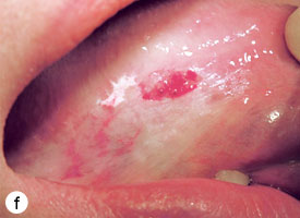
32 hours ago Biopsy Samples and the Diagnosis of Celiac Disease. Above: Normal villi; Below: Celiac Disease. Those who have the standard clinical symptoms of celiac disease, such as sensitivity to gluten, irritability, abdominal pain, or a positive blood test, may undergo an endoscopy to confirm their … >> Go To The Portal
How is small intestinal biopsy used to diagnose celiac disease?
Small intestinal biopsy. Small intestinal biopsy is the cornerstone of diagnosis and should be undertaken in all patients with suspected celiac disease. Biopsies can be obtained using a capsule with a suction-guillotine mechanism (e.g. Watson capsule).
How are biopsies used to diagnose cancer?
Most cancer patients will undergo a biopsy or other procedure to remove a sample of tissue for examination by a pathologist in order to diagnose their disease. There are a variety of methods used to obtain samples, including a typical biopsy, fine needle aspiration, or a biopsy with the use of an endoscope.
What are the characteristics of intestinal biopsies of colonic cancer?
Intestinal biopsies are characterized by severe villus atrophy, crypt hyperplasia, and a mixed inflammatory infiltrate in the lamina propria (see Figs. 9-6 and 9-7 ). Concomitant colitis and gastritis are present in the majority of cases.
How many biopsies are needed to diagnose a pyloric stenosis?
(2) One biopsy from the greater curvature of the antrum within 2–3 cm of the pylorus (3) One biopsy from the lesser curvature of the body 8 cm from the cardia (4) One biopsy from the greater curvature of the body 8 cm from the cardia
What do intestinal biopsies show?
The small-bowel biopsy is used to diagnose and confirm disease of the lining of the small intestine. Evaluation of patients with malabsorption, iron deficiency (anaemia), coeliac disease, neoplasia.
Will a biopsy show celiac disease?
Endoscopies and biopsies are the best way to diagnose celiac disease. A gastroenterologist (doctor who treats people with disorders of the stomach and intestines) will do an endoscopy if your/your child's blood tests or genetic tests show signs of celiac disease.
What tests confirm celiac disease?
Two blood tests can help diagnose it: Serology testing looks for antibodies in your blood. Elevated levels of certain antibody proteins indicate an immune reaction to gluten. Genetic testing for human leukocyte antigens (HLA-DQ2 and HLA-DQ8) can be used to rule out celiac disease.
Is biopsy necessary for celiac diagnosis?
The only way to confirm a celiac disease diagnosis is to have an intestinal biopsy. A pathologist will assign a Modified Marsh Type to the biopsy findings. A Type of 3 indicates symptomatic celiac disease. However, Types 1 and 2 may also indicate celiac disease.
What is a positive result for celiac disease?
All celiac disease blood tests require that you be on a gluten-containing diet to be accurate. The tTG-IgA test will be positive in about 93% of patients with celiac disease who are on a gluten-containing diet. This refers to the test's sensitivity, which measures how correctly it identifies those with the disease.
What is normal range for celiac?
Optimal Result: 0 - 3 U/mL, or 0.00 - 100.00 ug/g. A tissue transglutaminase IgA (tTg-IgA) test is used to help doctors diagnose celiac disease or to see how well people with the condition are doing.
Is celiac disease serious?
Celiac disease is a serious autoimmune disease that occurs in genetically predisposed people where the ingestion of gluten leads to damage in the small intestine. It is estimated to affect 1 in 100 people worldwide, but only about 30% are properly diagnosed.
How does a biopsy help doctors determine if a patient has celiac disease?
In individuals with celiac disease, gluten damages the villi and causes them to flatten. As a result, the body can't get the nutrients it needs, which leads to many of the health problems associated with celiac disease. With a biopsy, doctors can see if the villi are flattened.
What is the most accurate test for celiac disease?
The tTG-IgA test is the preferred celiac disease serologic test for most patients. Research suggests that the tTG-IgA test has a sensitivity of 78% to 100% and a specificity of 90% to 100%.
How accurate is celiac biopsy?
Study results. The new study included about 1400 adults of which 431, or 30 percent had TTG test results of at least 10 times the upper limit of normal. Of these, 424, or 98 percent, had severe enough intestinal damage on biopsy to be diagnosed with celiac disease.
Can celiac disease be cured?
There's no cure for celiac disease — but for most people, following a strict gluten-free diet can help manage symptoms and promote intestinal healing.
Is celiac visible in endoscopy?
Endoscopically visible hallmarks of celiac disease are scalloped duodenal folds, grooves and fissurations (Table 1). This contrasts with healthy tissue, which is covered with finger-like villi that provide a large surface area for nutrient uptake.
What is a small intestinal biopsy?
Small intestinal biopsy is the cornerstone of diagnosis and should be undertaken in all patients with suspected celiac disease. Biopsies can be obtained using a capsule with a suction-guillotine mechanism (e.g. Watson capsule).
What are the findings of a small bowel biopsy?
The striking finding is that the lamina propria is packed with macrophages, which have enlarged regions of cytoplasm with a foamy appearance. With periodic acid–Schiff (PAS) stain, the macrophages are stained a bright red due to the numerous bacteria contained within them. Some of these bacteria can be seen with electron microscopy to be dividing, a sign that the bacteria are alive and proliferating. In patients who are HIV positive and have the AIDS complex, PAS-positive macrophages can also be found due to the presence of Mycobacterium avium-intracellulare. These organisms can be distinguished from T. whippelii on electron microscopy because of differences in morphology.
What is the diagnosis of celiac disease?
Diagnosis/histology. A small bowel biopsy is obligatory to confirm a diagnosis of celiac disease. The histologic features include increased numbers of intraepithelial lymphocytes, villous shortening or flattening, crypt hyperplasia, and infiltration of the lamina propria with lymphoid cells.
Why is a small bowel biopsy important?
Small bowel biopsy is important in the evaluation of watery diarrhea. It can detect mucosal diseases such as celiac disease, previously discussed, in addition to eosinophilic enteritis, with the highest yield through duodenal biopsies. Infections with organisms such as Mycobacterium tuberculosis and Strongyloides stercoralis may be identified ...
What are the uncommon small bowel diseases that can be recognized on biopsy?
Uncommon small bowel diseases that can be recognized on biopsy include Whipple's disease, collagenous sprue, and lymphoma which have either secretory physiology or a combination of pathophysiological mechanisms to explain diarrhea. View chapter Purchase book. Read full chapter.
How many biopsies are required for gluten free diet?
The original criteria requiring a series of three biopsies, i.e., first to confirm the diagnosis, second for demonstration of response to a gluten-free diet, and the third for deterioration after gluten challenge, are required only in those few patients in which there still remains some diagnostic uncertainty.
Can a small bowel biopsy diagnose celiac sprue?
Based on the 1990 revised criteria of the European Society of Pediatric Gastroenterology and Nutrition, the diagnosis of celiac sprue can be made with a diagnostic small bowel biopsy in a patient with highly suggestive clinical symptoms, followed by an objective clinical response to a gluten-free diet.
What is a biopsy report?
A biopsy report describes the findings of a specimen. It contains the following information: Gross description. A gross description describes how it looks to the naked eye and where the biopsy was taken from. It may include a description of the color, size, and texture of the specimen. Microscopic exam.
How are histologic sections prepared?
Histologic sections are prepared in one of two ways: Permanent sections. The specimen is put into a fluid called a fixative for several hours, depending on the specimen type. The fixed speci men is put into a machine that removes the water from the speci men, and replaces it with paraffin wax. The paraffin-impregnated specimen is embedded ...
What is the machine that cuts thin sections of paraffin block containing the biopsy specimen?
A machine called a microtome cuts thin sections of the paraffin block containing the biopsy specimen. The sections are then placed on a glass slide and dipped into a series of stains or dyes to change the color of the tissue. The color makes cells more distinctive when viewed under a microscope. Frozen sections.
What is a smear on a slide?
Smears. Smears are done when the specimen is a liquid or there are small, solid chunks suspended in liquid. These are "smeared" onto a slide. They are then allowed to dry or are fixed. The fixed smears are stained, covered with a coverslip, and then examined under a microscope.
What is a microscopic exam?
A microscopic exam is a description of what the findings of the slides showed under a microscope. It's usually technical and not in simple language. Diagnosis. This is usually considered the "bottom line.".
Can a breast specimen be examined after it has been removed?
The specimen can be examined shortly after it has been removed from the patient. For example, surgical pathologists work closely with the surgeons during surgery for breast cancer. Often, a frozen section is used to determine how much of the breast tissue to remove. Smears.
Who prepares a written report for a biopsy?
After the biopsy specimen is obtained by the doctor, it is sent for examination to another doctor, the anatomical pathologist, who prepares a written report with information designed to help the primary doctor manage the patient’s condition properly.
What type of biopsy is used to remove a lump from the soft tissue?
2. Incisional biopsy. Only a portion of the lump is removed surgically. This type of biopsy is most commonly used for tumors of the soft tissues (muscle, fat, connective tissue) to distinguish benign conditions from malignant soft tissue tumors, called sarcomas. 3.
What is a colposcope?
The colposcope is actually a close- focusing telescope that allows the physician to see in detail abnormal areas on the cervix of the uterus, so that a good representation of the abnormal area can be removed and sent to the pathologist. 5. Fine needle aspiration. FNA biopsy.
What is the procedure called when you take a sample of tissue from a patient?
This procedure is called a biopsy , a Greek-derived word that may be loosely translated as “view of the living.”.
How many cells are drawn up in a syringe?
A needle no wider than that typically used to give routine injections (about 22 gauge) is inserted into a lump (tumor), and a few tens to thousands of cells are drawn up (aspirated) into a syringe. These are smeared on a slide, stained, and examined under a microscope by the pathologist.
Can you biopsy a thyroid?
Any organ in the body can be biopsied using a variety of techniques, some of which require major surgery (e.g., staging splenectomy for Hodgkin’s disease), while others do not even require local anesthesia (e.g., fine needle aspiration biopsy of thyroid, breast, lung, liver, etc).
Is intestinal metaplasia a risk factor for cancer?
For instance, a type of intestinal metaplasia of the stomach (in which columnar epithelium of the intestinal type replaces that of the gastric type) is considered a risk factor for the subsequent development of cancer of the stomach.
What is liquid biopsy?
What you need to know about liquid biopsies. A typical biopsy involves the surgical removal of a mass of abnormal cells. Fine needle aspiration involves guiding a thin needle into the cancer and gently sucking out cells for microscopic evaluation.
Why do doctors use liquid biopsies?
The method used to gain a tissue sample depends on the type of mass and location in the body. Doctors are increasingly using "liquid" biopsies to evaluate cancer which are easily collected from the blood and are non-invasive. Liquid biopsies are replacing the need to collect tissue in many situations.
What is the microscopic description of a tissue?
Microscopic Description: In the microscopic description, the pathologist describes how the cells of the tissue sample appear under a microscope. Specific attributes that the pathologist may look for and describe may include cell structure, tumor margins, vascular invasion, depth of invasion and pathologic stage.
What is the purpose of a pathologist's report?
The pathologist then writes a pathology report summarizing his or her findings.
What is the procedure to remove tissue from a cancer patient?
Most cancer patients will undergo a biopsy or other procedure to remove a sample of tissue for examination by a pathologist in order to diagnose their disease. There are a variety of methods used to obtain samples, including a typical biopsy, fine needle aspiration, or a biopsy with the use of an endoscope.
What is a pathologist?
A pathologist is a physician specializing in the diagnosis of disease based on examination of tissues and fluids removed from the body. Upon examination, the pathologist determines if the tissue sample contains normal, pre-cancerous or cancerous cells and then writes a report with his or her findings.
What is a histologic grade?
The histologic grade helps the pathologist identify the type of tumor. The grade may be described numerically with the Scarff-Bloom-Richardson system (1-3) or as well-differentiated, moderately-differentiated or poorly differentiated. Grade 1 or well-differentiated: Cells appear normal and are not growing rapidly.
How to tell if a biopsy is cancerous?
This is known as histologic (tissue) examination and is usually the best way to tell if cancer is present. The pathologist may also examine cytologic (cell) material.
What is the procedure to cut tissue into thin sections?
The tissue removed during a biopsy or surgery must be cut into thin sections, placed on slides, and stained with dyes before it can be examined under a microscope. Two methods are used to make the tissue firm enough to cut into thin sections: frozen sections and paraffin-embedded (permanent) sections.
What is an IHC report?
For example, the pathology report may include information obtained from immunochemical stains (IHC). IHC uses antibodies to identify specific antigens on the surface of cancer cells. IHC can often be used to: Determine where the cancer started.
How are tissue samples prepared?
All tissue samples are prepared as permanent sections, but sometimes frozen sections are also prepared. Permanent sections are prepared by placing the tissue in fixative (usually formalin) to preserve the tissue, processing it through additional solutions, and then placing it in paraffin wax.
How long does it take for a pathologist to send a report?
The pathologist sends a pathology report to the doctor within 10 days after the biopsy or surgery is performed. Pathology reports are written in technical medical language. Patients may want to ask their doctors to give them a copy of the pathology report and to explain the report to them. Patients also may wish to keep a copy ...
What is the procedure to remove tissue under a microscope?
In most cases, a doctor needs to do a biopsy or surgery to remove cells or tissues for examination under a microscope. Some common ways a biopsy can be done are as follows: A needle is used to withdraw tissue or fluid.
What is the purpose of endoscope?
An endoscope (a thin, lighted tube) is used to look at areas inside the body and remove cells or tissues. Surgery is used to remove part of the tumor or the entire tumor. If the entire tumor is removed, typically some normal tissue around the tumor is also removed. Tissue removed during a biopsy is sent to a pathology laboratory, ...
Popular Posts:
- 1. patient portal the doctors clinc
- 2. caroline kingston md santa fe patient portal
- 3. orange park medical center patient portal
- 4. lancaster county podiatry patient portal
- 5. columbia physicians patient portal
- 6. can you create a patient portal with pagecloud.com
- 7. orthopedic associaters fo port huron patient portal
- 8. french hospital create patient portal
- 9. north florida regional medical center patient portal login
- 10. breese il hospital patient portal