Sample Reports – National Diagnostic Imaging
11 hours ago · X-Ray #1 X-Ray #2 X-Ray #3 X-Ray #4 X-Ray #5 X-Ray Report Sample #1 HANDS AND WRISTS – TWO VIEWS: Hands and wrists, two views of the right and left hand and wrist were obtained. There is generalized osteopenia. There are OA changes seen at the first CMC joint with subchondral sclerosis and joint space narrowing. Ulnar styloids appear intact. >> Go To The Portal
Example MRI
Magnetic resonance imaging
Magnetic resonance imaging is a medical imaging technique used in radiology to form pictures of the anatomy and the physiological processes of the body. MRI scanners use strong magnetic fields, magnetic field gradients, and radio waves to generate images of the organs in the body. MRI does not involve X-rays or the use of ionizing radiation, which distinguishes it from CT or CAT scans and PET sca…
Full Answer
How to write a medical report for a patient?
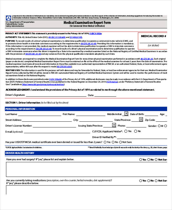
To write the report, it’s best to use proper wording that a reader may understand. Keep in mind that the people who may get a hold of the report may or may not be a part of the medical field. The report should contain a brief but understandable executive summary of the actual result.
What are the different types of medical reports?
What are the different kinds of medical reports? For some of the more in-depth and extensive examples, the different kinds of medical reports often include radiology reports, printable laboratory reports, and pathology reports. How does a medical report differ from a prescription?
Can a patient request a copy of a radiology report?
Patients may request copies of imaging studies and/or copies of their radiology reports. Radiology reports will be available to patients following the radiologist’s interpretation of the imaging study. Patients must complete a radiology patient health information release form before the report or images can be released to the patient.
When will my radiology report be available to patients?
Radiology reports will be available to patients following the radiologist’s interpretation of the imaging study. Patients must complete a radiology patient health information release form before the report or images can be released to the patient.

What are some examples of medical imaging?
Common types of imaging include:X-rays.CT (computed tomography) scan.MRI (magnetic resonance imaging)ultrasound.nuclear medicine imaging, including positron-emission tomography (PET)
What is an imaging report?
A radiology report includes complex anatomical and medical terms specifically written for healthcare providers. A radiologist (a physician specially trained in medical imaging) reviews your medical history and analyzes your diagnostic imaging. Next, the radiologist writes a report detailing the results.
How do you describe medical imaging?
Medical imaging, also known as radiology, is the field of medicine in which medical professionals recreate various images of parts of the body for diagnostic or treatment purposes. Medical imaging procedures include non-invasive tests that allow doctors to diagnose injuries and diseases without being intrusive.
What is an example of an imaging test?
Examples of imaging tests are computed tomography (CT), mammography, ultrasonography, magnetic resonance imaging (MRI), and nuclear medicine tests. Also called imaging procedure.
How do you write a radiology report?
10:4028:23Featured Video - How to make a great radiology report - YouTubeYouTubeStart of suggested clipEnd of suggested clipSo the emphasis and findings is on short informative and factual phrases. And the impression is theMoreSo the emphasis and findings is on short informative and factual phrases. And the impression is the meaning of the findings which leads to a diagnosis or differential diagnosis.
How do I read my MRI report?
MRI interpretation Systematic approachStart by checking the patient and image details.Look at all the available image planes.Compare the fat-sensitive with the water-sensitive images looking for abnormal signal.Correlate the MRI appearances with available previous imaging.Relate your findings to the clinical question.
What are the 4 types of medical imaging?
What Are The Different Types Of Medical Imaging?MRI. An MRI, or magnetic resonance imaging, is a painless way that medical professionals can look inside the body to see your organs and other body tissues. ... CT Scan. ... PET/CT. ... Ultrasound. ... X-Ray. ... Arthrogram. ... Myelogram. ... Women's Imaging.
What does a medical imaging study reveal?
Medical imaging seeks to reveal internal structures hidden by the skin and bones, as well as to diagnose and treat disease. Medical imaging also establishes a database of normal anatomy and physiology to make it possible to identify abnormalities.
What are the 5 types of medical imaging exams?
Learn more about our five most common modalities for our various types of imaging tests: X-ray, CT, MRI, ultrasound, and PET.
What is this medical image?
Medical imaging refers to several different technologies that are used to view the human body in order to diagnose, monitor, or treat medical conditions.
What is the most common form of medical imaging?
X-rays (radiographs) are the most common and widely available diagnostic imaging technique. Even if you also need more sophisticated tests, you will probably get an x-ray first.
What are different types of imaging tests?
Types of imaging testsComputed tomography (CT) scan.Magnetic resonance imaging (MRI) scan.Breast MRI.X-rays and other radiographic tests.Mammography.Nuclear medicine scans (bone scans, PET scans, Thyroid scans, MUGA scans, gallium scans)Ultrasound.
What are common inclusions in a medical report?
Besides the patient’s personal data, there are also multiple kinds of information written into these reports. Among the numerous inclusions would b...
What are the different kinds of medical reports?
For some of the more in-depth and extensive examples, the different kinds of medical reports often include radiology reports, printable laboratory...
How does a medical report differ from a prescription?
A medical report tends to be all-encompassing, complete with details of a patient’s illness and even prescriptions. If you’re just talking about pr...
Lumbar Spine MRI With and Without Contrast
Aggressive lesion of bone with invasion of the central canal and paraspinal soft tissues.
Shoulder MRI Report
Multiple rotator cuff tears and gross enlargement of the long head of the biceps tendon sheath.
What are some examples of medical reports?
For some of the more in-depth and extensive examples, the different kinds of medical reports often include radiology reports, printable laboratory reports, and pathology reports.
Why is it important to use professional language in medical reports?
Use professional language and ensure that there is enough clarity to prevent any misunderstandings among all of the involved parties.
Can you keep a copy of a medical report?
The creation of a medical report may dictate that you keep a separate but identical copy for yourself. The purpose of doing so is purely related to documentation. Also, in the event that the original medical report is somehow lost or tampered with, the patient can always turn back to you for references.
Is a medical report vague?
A medical report that comes off as vague is practically useless. For it to be valid and useful, the medical professional writing it must go into detail. With that said, use specific terms and provide particular comments and suggestions for the benefit of the report’s recipient.
Can you request a copy of a radiology report?
Patients may request copies of imaging studies and/or copies of their radiology reports. Radiology reports will be available to patients following the radiologist’s interpretation of the imaging study. Patients must complete a radiology patient health information release form before the report or images can be released to the patient.
Do you have to complete a release form for radiology?
Patients must complete a radiology patient health information release form before the report or images can be released to the patient. The release form is not necessary for the physician who ordered the exam to receive the report. Frequently Asked Questions.
What does it mean when a radiologist looks at an area of the body?
Sometimes an exam covers an area of the body but does not discuss any findings. This usually means that the radiologist looked but did not find any problems to tell your doctor.
What is a comparison in radiology?
Comparison. Sometimes, the radiologist will compare the new imaging exam with any available previous exams. If so, the doctor will list them here. Comparisons usually involve exams of the same body area and exam type. Example: Comparison is made to a CT scan of the abdomen and pelvis performed August 24, 2013.
What is a biopsy?
biopsy. combining the finding with clinical symptoms or laboratory test results. comparing the finding with prior imaging studies not available when your radiologist looked at your images. For a potentially abnormal finding, the radiologist may make any of the above recommendations.
What is a radiologist?
A radiologist is a doctor who supervises these exams, reads and interprets the images, and writes a report for your doctor. This report may contain complex words and information. If you have any questions, be sure to talk to your doctor ...
Why is it important to have access to your health records?
Online access to your health records may help you make more informed decisions about your healthcare. In addition, online access lets you share your radiology reports with other doctors electronically. This may increase the safety, quality, and efficiency of your care. top of page.
Can you read a radiologist's electronic health records?
Typically, the report is sent to this doctor, who then delivers the results to you. Many patients can read their electronic health records online. Sometimes, these records include radiology reports.
Abstract
In acute ischemic stroke, diffusion weighted imaging (DWI) shows hyperintensities and is considered to indicate irreversibly damaged tissue. We present the case of a young stroke patient with unusual variability in the development of signal intensities within the same vessel territory.
Background
Acute cerebral ischemia can be visualized with MRI diffusion weighted imaging (DWI) within minutes after its onset [ 1 ]. It has been suggested that in the majority of human stroke patients DWI positive cerebral ischemia indicate irreversible tissue damage leading to infarction [ 2 ].
Case presentation
A 35 year old female patient was admitted with incomplete global aphasia and a fluctuating slight hypesthesia of the left hand. Her baseline National Institutes of Health Stroke Scale score (NIHSS) was 4 and arterial hypertension was the only vascular risk factor.
Conclusions
We demonstrated that the degree and time course of DWI hyperintensities can differ within one lesion in the first day after stroke. To the best of our knowledge, this is the first time that the exact time course of DWI reversal and reappearance was visualized using highly repetitive MRI examinations.
Acknowledgements
This research received funding from the Federal Ministry of Education and Research via the grant Center for Stroke Research Berlin (01EO0801 and 01EO1301).
Rights and permissions
Open Access This article is distributed under the terms of the Creative Commons Attribution 4.0 International License ( http://creativecommons.org/licenses/by/4.0/ ), which permits unrestricted use, distribution, and reproduction in any medium, provided you give appropriate credit to the original author (s) and the source, provide a link to the Creative Commons license, and indicate if changes were made.
What is a patient case study?
Writing Your Patient Case Study. Since patient case studies are generally descriptive, they are under the a phenomenological principle. This means that subjectivity is entertained and allowed in research design. The medical scenarios are open to the researcher’s interpretation and input of insights.
How do case studies make a difference in the medical arena?
Patient case studies make a difference in the medical arena by reporting clinical interactions that can improve medical practices, suggest new health projects, as well as provide a new research direction. By looking at an event as it exists in the natural setting, case studies shed understanding on a complex medical phenomenon.
What is a case study?
Case studies are a qualitative research method that offers a complete and in-depth look into some of the situations that baffled medical science. They document the cases that escape the ordinary in a hospital that has seen a manifold of plights. They serve as cautionary tales of the intricacy in dealing with human health.
Why do medical practitioners use case studies?
Medical practitioners use case studies to examine a medical condition in the context of a research question. They perform research and analyses that adhere to the scientific method of investigation and abide by ethical research protocols. The following are case study samples and guides on case presentation.
Can you generalize a population using one case study?
You cannot generalize a population using one case study. However, multiple case study contains two or more cases under the point of interest can give you a replicated result. When the findings remain true for several cases under this research method, your case study’s results become more reliable.
Should you look into all possible explanations for a medical condition?
You should look into all of the possible explanations for the medical condition at hand. If a plight can be explained by more than one reason , then you have to look into the less obvious but similarly compelling explanations. Make your case study as informative as possible.
Is a clinical interaction report valid for generalization?
Since it documents stand-out clinical interactions where a single person or a few number of people are a party of, the findings may not be valid for generalization for a wider population.

Popular Posts:
- 1. providers direct patient portal
- 2. texas family medicine patient portal
- 3. white labeled telehealth patient portal
- 4. lexington infectious disease patient portal
- 5. patient portal southern medical
- 6. anderson hospital myhealth patient portal
- 7. jefferson employee portal
- 8. opa patient portal
- 9. ridgecrest regional hospital/patient portal
- 10. what are the minimum fields needed for patient signup?