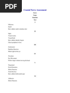
Cranial Nerves Chart & Assessment Cheat Sheet (2020)
6 hours ago •20/20 =s patient can read at 20` with same accuracy as person with normal vision. •20/400 =s patient can read @ 20` what normal person can read from 400` (i.e. very poor acuity). •If patient can’t identify all items correctly, number missed is listed after a ‘-’ sign (e.g. 20/80 … >> Go To The Portal
What should I expect during a cranial nerve examination?
There are many symptoms that could prompt a referral to a neurologist, including:
- Severe headaches
- Dizziness
- Prolonged numbness or tingling or numbness on one side of the body
- Chronic pain, including pain with weakness or numbness
- Seizures
- Intense muscle weakness
- Problems walking
- Tremors that affect daily activities
- Certain vision problems
- Changes in your personality
How to test all 12 cranial nerves?
Cranial nerve examination frequently appears in OSCEs. You’ll be expected to assess a subset of the twelve cranial nerves and identify abnormalities using your clinical skills. This cranial nerve examination OSCE guide provides a clear step-by-step approach to examining the cranial nerves, with an included video demonstration.
How to assess the cranial nerves?
- Pupils should be round and bilaterally equal in size. The diameter of the pupils usually ranges from two to five millimeters. ...
- Test pupillary reaction to light. ...
- Test eye convergence and accommodation. ...
- The acronym PERRLA is commonly used in medical documentation and refers to, “pupils are equal, round and reactive to light and accommodation.”
What are the 12 cranial nerve tests?
Nystagmus secondary to BPPV has the following nearly pathognomic characteristics:
- A latency period of 5 to 10 sec
- Usually, vertical (upward-beating) nystagmus when the eyes are turned away from the affected ear and rotary nystagmus when the eyes are turned toward the affected ear
- Nystagmus that fatigues when the Dix-Hallpike maneuver is repeated
How do I report a normal cranial nerve exam?
Documentation of a basic, normal neuro exam should look something along the lines of the following: The patient is alert and oriented to person, place, and time with normal speech. No motor deficits are noted, with muscle strength 5/5 bilaterally. Sensation is intact bilaterally.
How do you assess cranial nerve exam?
3:539:34How to do the Cranial Nerve Examination - YouTubeYouTubeStart of suggested clipEnd of suggested clipInclude forehead cheek and chin bilaterally. Ask the patient whether the same stimulus feels theMoreInclude forehead cheek and chin bilaterally. Ask the patient whether the same stimulus feels the same on both sides. Equal. Equal test cold sensation using the metal tuning fork.
What is a normal neurological examination?
The neurologic examination is typically divided into eight components: mental status; skull, spine and meninges; cranial nerves; motor examination; sensory examination; coordination; reflexes; and gait and station. The mental status is an extremely important part of the neurologic examination that is often overlooked.
What are the 7 areas of documentation of the neurological exam?
The neurological exam can be organized into 7 categories: (1) mental status, (2) cranial nerves, (3) motor system, (4) reflexes, (5) sensory system, (6) coordination, and (7) station and gait. You should approach the exam systematically and establish a routine so as not to leave anything out.
How do you check cranial nerve 9 and 10?
Cranial Nerves 9 & 10 - Motor The motor division of CN 9 & 10 is tested by having the patient say "ah" or "kah". The palate should rise symmetrically and there should be little nasal air escape. With unilateral weakness the uvula will deviate toward the normal side because that side of the palate is pulled up higher.
Why do nurses assess cranial nerves?
Assessment of the cranial nerves provides insightful and vital information about the patient's nervous system.
How do you write a good history of present illness?
The HPI should be written in prose with full sentences and be a narrative that builds an argument for the reason the patient was admitted.Has a starting point (i.e. “the patient was in her usual state of health until 5 days prior to admission.).Has appropriate flow, continuity, sequence, and chronologic order.More items...
What are the 4 components of a neurological check?
There are many components to a neurological exam, including cognitive testing, motor strength and control, sensory function, gait (walking), cranial nerve testing, and balance.
What is the most reliable indicator of a patient's neurologic function?
A patient's mental status is the most reliable indicator of brain function, so when there is altered mental status, obtaining a history and assessing for cerebellar function, weakness and paresthesia becomes far more difficult.
How do you document Heent?
Documenting a normal exam of the head, eyes, ears, nose and throat should look something along the lines of the following: Head – The head is normocephalic and atraumatic without tenderness, visible or palpable masses, depressions, or scarring. Hair is of normal texture and evenly distributed.
How do nurses do neurological assessments?
2:387:26Routine Neurological Assessments- Nursing Skills - YouTubeYouTubeStart of suggested clipEnd of suggested clipAnd make sure you ask them if there's any tenderness or pain. Then you can just lightly touch bothMoreAnd make sure you ask them if there's any tenderness or pain. Then you can just lightly touch both sides of their forehead cheeks and chin. And make sure the patient feels it equally on both sides.
What is a neurological exam?
The neurological exam consists of a number of components that assess for neurological abnormalities. The level of detail of the neurological exam performed in the clinical setting varies with each patient depending on history and symptoms. Patients presenting with neurological deficits, or symptoms of neurological conditions, for example, ...
What is neuro exam?
A neuro exam is one of the more complex body systems to master when it comes to assessment and documentation. Testing the cranial nerves, for example, takes practice. Omitting a small part of the process can mean missing a potentially serious diagnosis.
Do you need a neurological assessment?
Patients presenting with neurological deficits, or symptoms of neurological conditions, for example, may require a complete neurological assessment. Patients presenting for non-neurological complaints may only require a simple assessment of mental status.
Why wouldn't cranial nerves work?
Why wouldn’t a cranial nerve “work ”? In many neuro diseases, the neurons that supply a particular nerve is damaged, which makes the nerve not function properly. For example, in multiple sclerosis the myelin sheath of the neurons in the central nervous system are damaged, which leads to some sensory and motor problems.
What nerve causes blurry vision?
Therefore, you can assess this nerve (cranial nerve II) for any type of abnormalities.
How far can a patient see with normal vision?
This means the patient can see at 20 feet what a person with normal vision can see at 30 feet.
Which nerve is responsible for mastication?
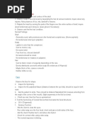
Cranial Nerve V. To test Cranial Nerve V…..trigeminal nerve: This nerve is responsible for many functions and mastication is one of them. Have the patient bite down and feel the masseter muscle and temporal muscle. Then have the patient try to open the mouth against resistance.
How to test eye movement?
Test eye movement by using a penlight. Stand 1 foot in front of the patient and ask them to follow the direction of the penlight with only their eyes. At eye level, move the penlight left to right, right to left, up and down, upper right to lower left, and upper left to lower right.
How to test each ear individually?
Each ear is tested individually. The patient should be instructed to occlude the non-test ear with their finger. Exhale before whispering and use as quiet a voice as possible. Whisper a combination of numbers and letters (for example, 4-K-2), and then ask the patient to repeat the sequence.
How to test sternocleidomastoid muscle?
Test the right sternocleidomastoid muscle. Face the patient and place your right palm laterally on the patient’s left cheek. Ask the patient to turn their head to the left while resisting the pressure you are exerting in the opposite direction. At the same time, observe and palpate the right sternocleidomastoid with your left hand. Then reverse the procedure to test the left sternocleidomastoid.
What does a positive Romberg test mean?
It is expected that the patient will maintain balance and stand erect. A positive Romberg test occurs if the patient sways or is unable to maintain balance. The Romberg test is also a test of the body’s sense of positioning (proprioception), which requires healthy functioning of the spinal cord.
How to test cranial nerves II and III?
The pupillary light reflex tests both cranial nerves II and III. First, inspect both pupils and make sure they are equal in size and shape. Then dim the lights if possible and shine a penlight directly into the right eye. Both pupils should constrict and maintain symmetry. Note if they are brisk or sluggish and if they are symmetric. Remove the light source and watch both eyes dilate equally as well. Do the same for the left eye.
Why is cranial nerve assessment important?
The cranial nerve assessment is an important part of the neurologic exam, as cranial nerves can often correlate with serious neurologic pathology. This is important for nurses, nurse practitioners, and other medical professionals to know how to test cranial nerves and what cranial nerve assessment abnormalities may indicate.
What nerve controls the eyeball?
The fourth cranial nerve, the trochlear nerve, innervates the superior oblique muscle of the eyes. This means it controls the downward movement of the eyeball and prevents it from rolling upward. When there is a fourth nerve palsy, patients will often complain of vertical diplopia and/or tilting of objects.
What nerve controls the movement of the eyelid?
The oculomotor nerve controls the majority of the extraocular muscles. It is primarily responsible for eye movement, eyelid movement, and pupillary constriction. If there is any oculomotor nerve impairment, there will be a pupillary dilation, ptosis ( drooping eyelid ), and outward deviation of the eye – termed abduction. When a patient has diplopia (double vision), it is often due to a unilateral lesion on this cranial nerve. In most cases, third nerve palsy resolves over weeks to months.
What nerves are tested for oculomotor nerves?
To test the oculomotor nerve, you need to assess the EOMs. Testing the EOMs also tests cranial nerves IV and VI, as all three nerves are responsible for eye movement.
What nerve is responsible for the sense of smell?
The olfactory nerve is responsible for the sense of smell. Although rarely tested in practice, alterations in smell can be caused by serious intracranial pathology (brain tumors, strokes, TBI), neurodegenerative diseases like Alzheimer’s, Parkinson’s, or MS, or benign and transient causes such as the common cold.
How to test hypoglossal nerve?
How to test the Hypoglossal Nerve. To test the hypoglossal nerve, ask the patient to stick out their tongue. If the tongue deviates to one side , this indicates hypoglossal nerve dysfunction on the side of deviation. Then ask them to move their tongue from side to side rapidly.
Popular Posts:
- 1. san antonio urology patient portal
- 2. online patient portal tricare
- 3. partners patient gateway - login
- 4. baptist north patient portal
- 5. archconnect patient portal
- 6. patient portal + uma chatterjee
- 7. patient portal mountain star
- 8. wile-cornell patient portal
- 9. st. luke hospital phillipsburg patient portal
- 10. dr paz patient portal